CUHK
News Centre
CUHK Researcher Uncovers Mechanism Explaining Why Fathers’ Mitochondrial DNA is Not Inherited to Offspring
Prof. Byung-Ho Kang, an Associate Professor of the School of Life Sciences at The Chinese University of Hong Kong (CUHK) and researchers at the University of Colorado have uncovered novel aspects of the mystery of maternal inheritance of mitochondrial DNA through a multi-disciplinary approach. The findings have recently been published in Science, one of the most prestigious and authoritative scientific journals*.
Some 50 years ago, scientists discovered that our DNA does not only reside in the nucleus of our cells, but also in structures called mitochondria, the power plant of our cells. Mitochondrial DNA accounts for only a small proportion of the total DNA in the human body, encoding for only 37 genes out of an estimate total of 20,000 genes. In most animal species, mitochondrial DNA is inherited solely from the mother, unlike the nuclear DNA that is inherited from both parents. Why and how fathers’ mitochondrial DNA disappears from the zygotes remains a mystery to biologists.
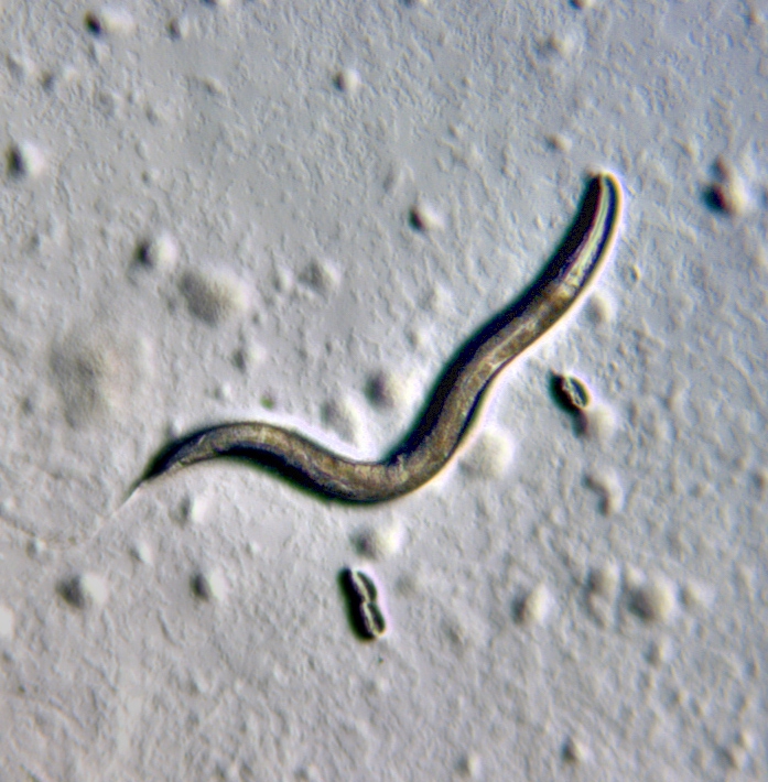
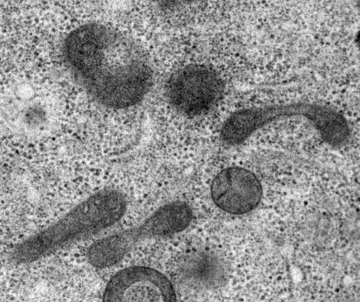
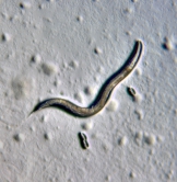
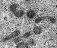
To solve the mystery, Professor Kang at CUHK and Prof. Ding Xue’s group at the University of Colorado Boulder examined the mitochondria in the sperm of a roundworm called C. elegans using electron tomography that reveals cells’ inner structures at nanometer-level resolutions in three-dimension. They observed that sperm mitochondria in C. elegans exhibit signs of decay before the sperm reaches any autophagosome, a cell structure known to engulf sperm mitochondria in a process named autophagy in the eggs of the organism.
Professor Kang said, ‘We found that the sperm mitochondria started self-degradation as if they were committing suicide. This internal self-destruct mechanism gets activated once a sperm penetrates an egg. We also found that delaying this mechanism will lower the rates of embryo survival. This may help scientists understand diseases related to this mechanism and improve in vitro fertilization techniques.’
The research team then searched for mitochondrial proteins that contribute to the self-initiated elimination of sperm mitochondria. They found that CPS-6 localized on the surface of sperm mitochondria can chop off DNA molecules. These results provide a direct molecular explanation of how paternal mitochondrial DNA is erased in offspring. They also showed that paternal mitochondria could do harm in developing C. elegans embryos if they are not removed.
Mitochondria are often damaged when they produce cells’ energy. They are under continuous surveillance and abnormal mitochondria are removed by autophagy. If this cleaning job becomes aberrant in human, one can suffer from neurodegenerative diseases. Further research on paternal mitochondrial elimination in C. elegans could provide new insight into autophagy in human cells and how to devise cures for diseases stemming from problems in autophagy, including Parkinson’s disease, dementia, some forms of heart diseases and blindness.
Professor Kang remarked, ‘This research could not have been possible without the combined use of electron microscopy imaging and extensive searching for genes involved in the degradation of paternal mitochondria. Adopting multi-disciplinary approaches is critical for tackling difficult questions in biology. The success of this research exemplifies the importance of collaboration between laboratories specializing in different techniques. I would like to thank the Hong Kong Research Grants Council for supporting the Centre for Organelle Biogenesis and Function at CUHK under the Areas of Excellence Scheme which greatly facilitates the electron microscopy investigation of sperm mitochondria in this project.’
About Prof. Byung-Ho Kang
Prof. Byung-Ho Kang is currently an Associate Professor of the School of Life Sciences at CUHK and a member of the multi-disciplinary research Center for Organelle Biogenesis and Function funded and supported by the Areas of Excellence Scheme of the Hong Kong Research Grants Council (AoE/M-05/12). He obtained his BSc in Chemistry from Seoul National University and a PhD in Biochemistry from the University of Wisconsin-Madison. Before joining CUHK, he was an associate professor at the Department of Microbiology and Cell Sciences of the University of Florida.
Professor Kang is an expert in cell biology and advanced electron microscopy. He and his team are studying organelle biogenesis in animal and plant cells, combining theuse of molecular biology and advanced electron microscopy techniques. His research on C. elegans has yielded novel and important information on apoptosis and autophagy. Professor Kang has published in top international scientific journals, including Science, Molecular Cell, Current Biology, Proceedings of the National Academy of Sciences of the United States of America, The Plant Cell, and Plant Physiology.
Please visit the website of CUHK Center for Organelle Biogenesis and Function:
http://www.cuhk.edu.hk/centre/iCell/
*ORIGINAL ARTICLE: Mitochondrial endonuclease G mediated breakdown of paternal mitochondria upon fertilization. Science http://dx.doi.org/10.1126/science.aaf4777 (2016)
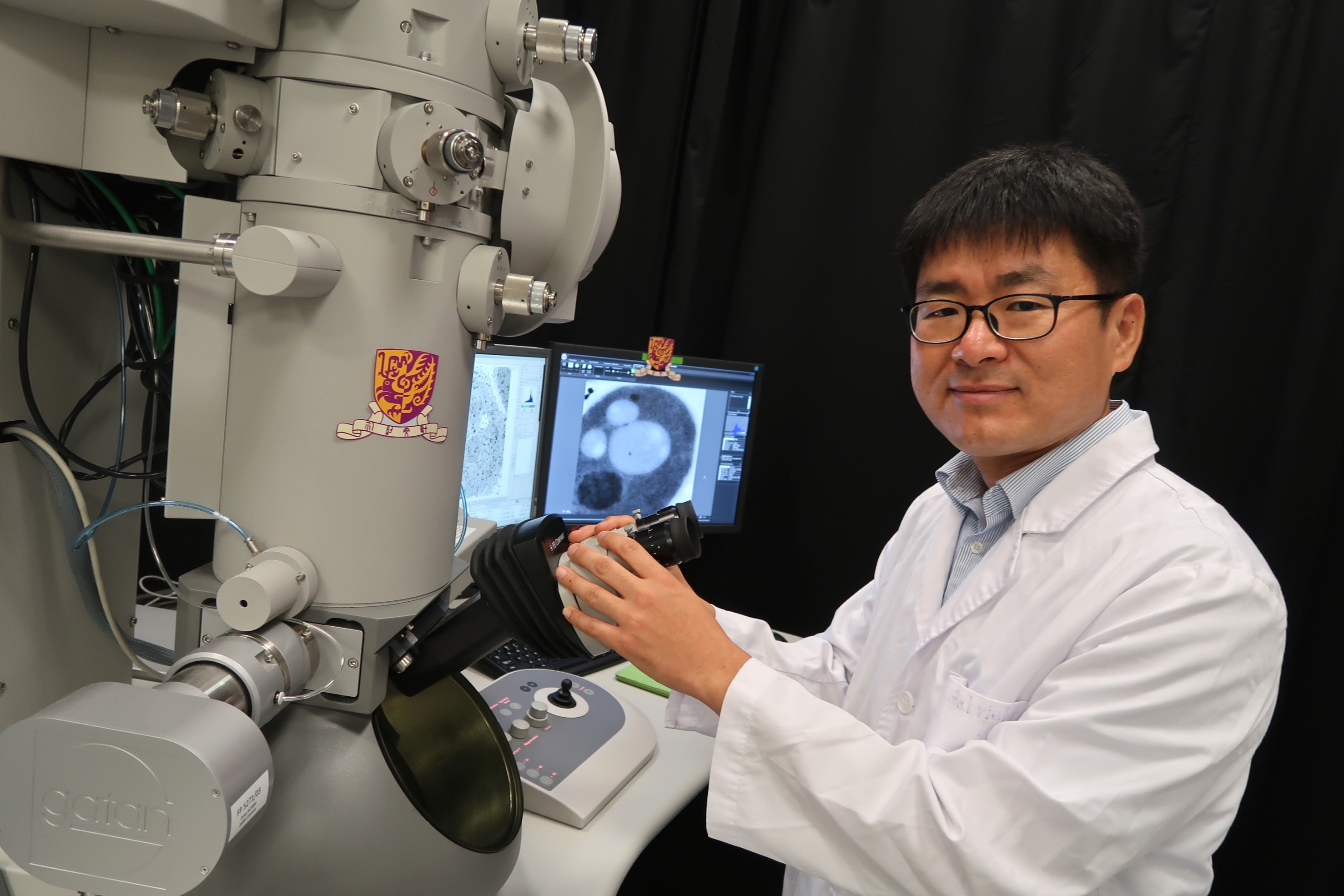
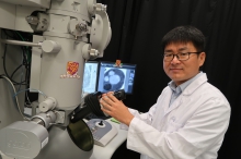
Prof. Byung-Ho Kang reveals the self-degradation of sperm mitochondria in C. elegans using electron tomography. The results are published in Science.
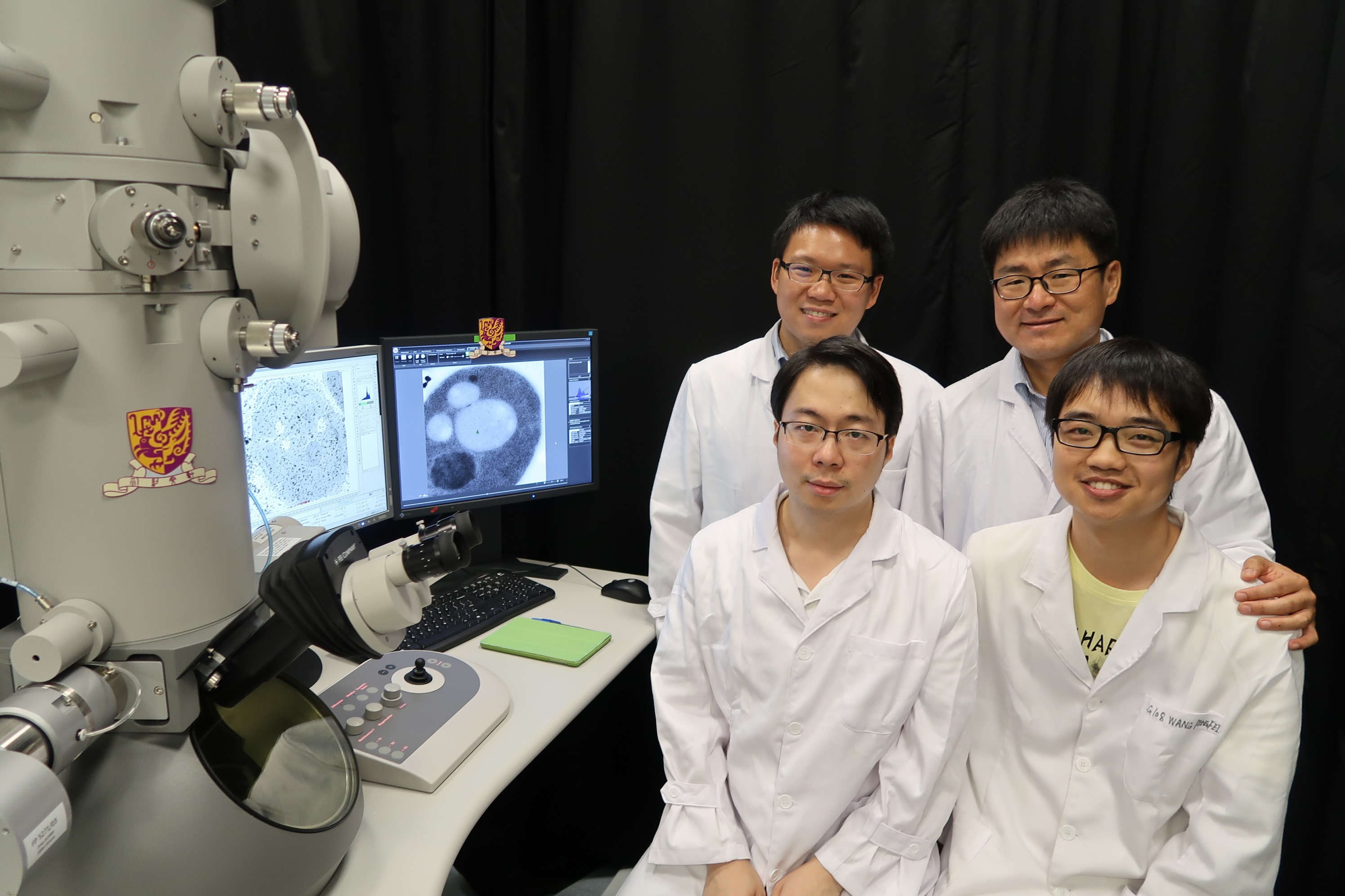
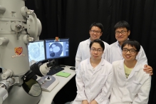
Prof. Byung-Ho Kang and his research team members: Keith Ka Ki Mai, Pengfei Wang and Dr Zizhen Liang
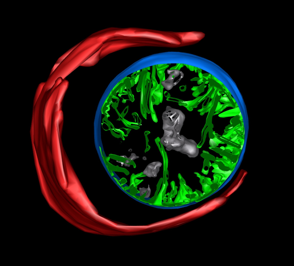
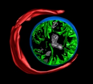
3-D model of maternal autophagosome (red) engulfing a paternal mitochondrion (blue), generated from electron tomography data


