CUHK
News Centre
CUHK is the First in Asia to Introduce the Real-Time 3-Dimensional Transesophageal Echocardiography to Diagnose Heart Diseases
Ultrasound of the heart, or echocardiography, is the most commonly used non-invasive imaging test by cardiologists. It helps to diagnose various heart diseases, guide therapy and monitor treatment progress. One of the particularly useful tools is to perform ultrasound examination of the heart by passing an endoscope into the esophagus, which is called transesophageal echocardiography (TEE). Although it has improved the quality of ultrasound images and provided doctors with additional information on the structure of the heart, such as the heart valves problem and the presence of atrial septal defect, this examination has major limitations. As the current TEE technology only provides two-dimensional ultrasound images, i.e. only a cut-plane of the heart at a time, it is unable to provide comprehensive information on the structure and function of the heart which is a three-dimensional organ. On the other hand, accurate and detailed information is very important for cardiologists to estimate disease risk and to plan for the best treatment regimen.
In March 2008, Professor Yu Cheuk-man and Professor Yip Wai-kwok from the Division of Cardiology of Department of Medicine and Therapeutics, and the Institute of Vascular Medicine of The Chinese University of Hong Kong (CUHK) were the first in Asia to introduce a new ultrasound technology to Hong Kong, namely the real-time three-dimensional TEE, or 3D-TEE. The 3D-TEE displays the true three-dimensional structure instantaneously at real-time. Doctors can rotate the heart image to any preferred angle or zoom into structures of clinical interest. The scanning time is also reduced. Since the introduction of the 3D-TEE, CUHK has examined 142 patients in the Prince of Wales Hospital, including 58 patients with valvular heart disease, 33 patients with congenital heart disease and 51 patients with other diagnoses such as suspected endocarditis, searching for cardiac causes of stroke and guidance for catheter ablation procedures of cardiac arrhythmias.
The 3D-TEE is found to have the following unique advantages. Firstly, this new tool is more accurate in assessing the severity of valvular heart disease such as leakage or narrowing of valves, and permits accurate measurement of the size and shape of congenital defects between the atria or ventricles. Secondly, the excellent image quality of 3D-TEE can help cardiac surgeons to plan and decide the type of valve surgery in advance, as cardiologists are able to, for the first time, examine the image of the valve identical to what surgeons will encounter during surgery. Lastly, this new technology is very useful in guiding catheter-based treatment procedures in cardiology such as closure of congenital heart defects and ablation of cardiac arrhythmias.
To share the first-hand experiences with other experts in Asia, the Division of Cardiology and the Institute of Vascular Medicine of CUHK hosts the “First 3D Matrix TEE Conference with Live Scanning and Interactive Imaging Sessions” on 28 and 29 April at the Prince of Wales Hospital. As the first conference of its kind in Asia, it attracts experts from China, Taiwan, Korea, Singapore, Malaysia and Thailand. The conference provides live demonstration on how 3D-TEE is performed, lectures and case-based workshops.
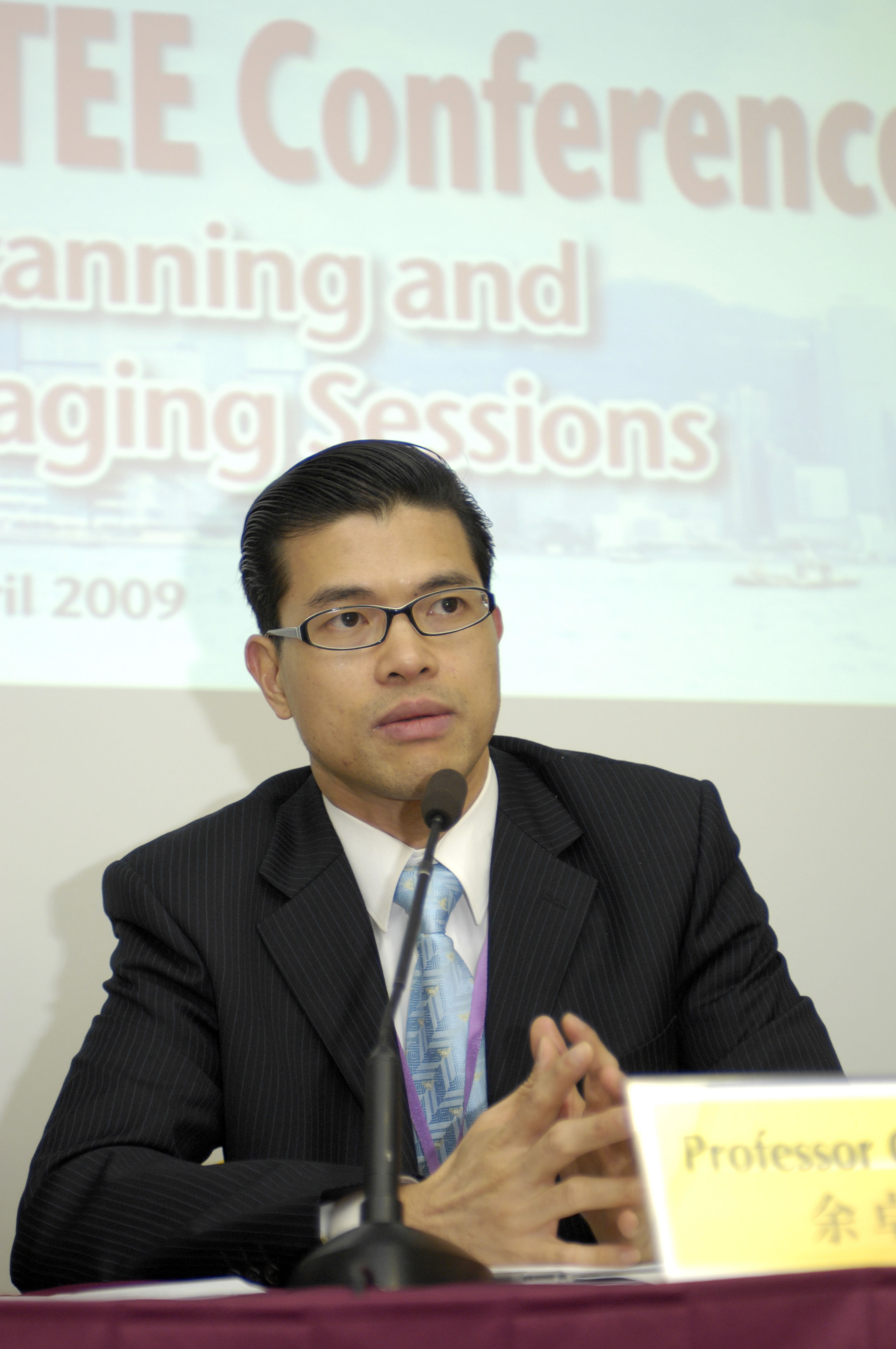
Professor Cheuk Man YU, Professor of Medicine & Head of Division of Cardiology, Department of Medicine & Therapeutics, Faculty of Medicine Director (Clinical Science), Institute of Vascular Medicine, CUHK
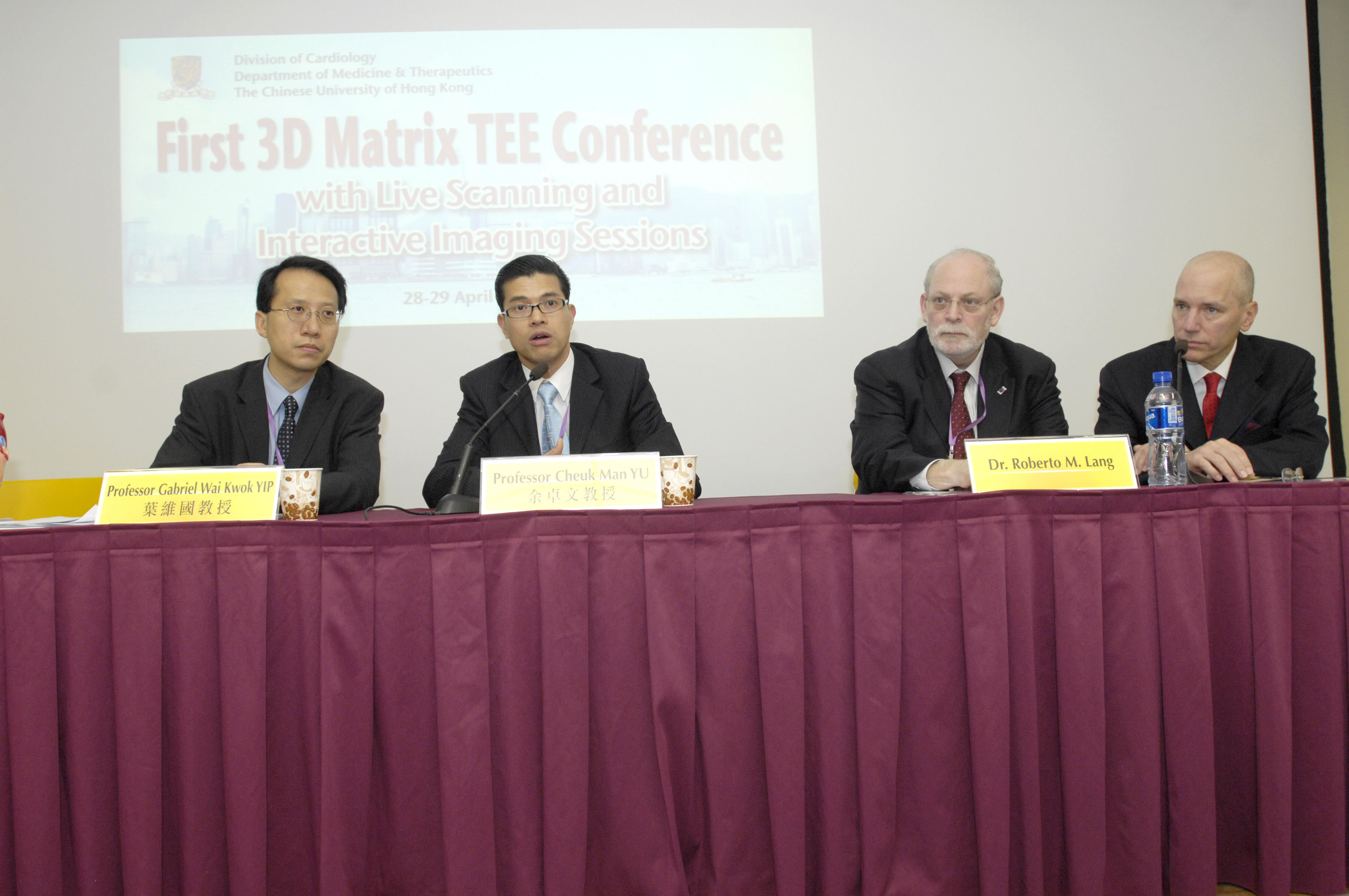
(From left) Professor Gabriel Wai Kwok YIP, Associate Professor, Division of Cardiology, Department of Medicine & Therapeutics, Faculty of Medicine, CUHK; Professor Cheuk Man YU, Professor of Medicine & Head of Division of Cardiology, Department of Medicine & Therapeutics, Faculty of Medicine Director (Clinical Science), Institute of Vascular Medicine, CUHK; Dr. David H. ADAMS, Marie-Josée and Henry R. Kravis Professor and Chairman, Department of Cardiothoracic Surgery, Mount Sinai Medical Center, New York, USA; and Dr. Roberto M. LANG, Professor of Medicine, Director, Noninvasive Cardiac Imaging Laboratories, Associate Director, Cardiology Fellowship Program, The University of Chicago Medical Center, Chicago, USA


