CUHK
News Centre
CUHK and University of Edinburgh palaeobiologists solve tail function mystery in the earliest flying vertebrates
Revealing never-seen-before soft tissue details using laser imaging
Pterosaurs were the earliest flying vertebrates and the first with membranous wings. These reptiles ruled the skies for over 150 million years during the Age of Dinosaurs around 225 to 66 million years ago, long before the emergence of birds and bats. Dr Michael Pittman, Assistant Professor at The Chinese University of Hong Kong (CUHK)’s School of Life Sciences, and Dr Natalia Jagielska of the Department of GeoSciences at the University of Edinburgh, led an international team that recently made progress in resolving the longstanding mystery of how pterosaurs achieved this success. Published in the prestigious journal eLife, their study clarifies the function of the tail in early pterosaurs using never-before-seen soft tissue details uncovered with laser imaging techniques.
Early pterosaurs had a long, bony tail that sometimes ended with a mysterious soft tissue tail “vane”. Unfortunately, knowledge of the structure of the tail vane had been insufficient to understand how it was used, especially since no living flying vertebrates have a similar tail to compare it with. Although the fossil record of ancient vertebrates is usually restricted to fossilised bones, in rare instances, delicate elements such as skin can be preserved. Using lasers, it is possible to study even more of these precious details, bringing ancient animals back to life in totally unexpected ways.
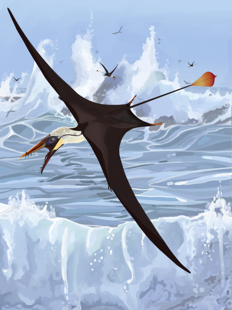
Through a screening process involving over 100 pterosaur fossils from across the world, Dr Pittman and his colleague Mr Thomas G Kaye of the Foundation for Scientific Advancement identified four specimens of Rhamphorhynchus that preserve otherwise unknown details of the tail vane. Rhamphorhynchus lived in the Late Jurassic, about 150 million years ago. It was no bigger than a seagull and had jaws filled with needle-like teeth. The team, including members from Hong Kong, Poland, the United States and Japan, visited the National Museum of Scotland in Edinburgh, the Natural History Museum in London and the Royal Ontario Museum in Toronto to study these four exceptional fossils.
Innovative laser imaging uncovers the key to pterosaurs’ flight success
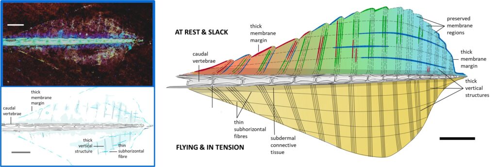
The vertically orientated tail vane was known to be supported by thick, vertical structures, but it was unclear what would have stopped it from fluttering like a flag in the wind, a tendency that would have created substantial unwanted drag, negatively impacting flight performance. Using lasers to make the specimens glow in the dark, a method called Laser-Stimulated Fluorescence, the team revealed that that the tail vane was in fact stiffened by a lattice formed by the thicker, hollow vertical structures cross-linking with thinner, subhorizontal fibres that ran to the tail tip. In doing so, the team uncovered a key innovation that paved the way for pterosaurs to conquer the skies.
Dr Nick Fraser of the National Museums Scotland, who was not involved in the study, said: “Without the researchers’ vision to apply new technology to apparently well-understood fossils, this tail vane would have remained in the dark. It is exciting to now see a critical feature of the pterosaur’s anatomy so beautifully displayed.”
First author Dr Natalia Jagielska, who will join CUHK in early 2025 as a Postdoctoral Research Fellow, added: “It never ceases to astound me that, despite hundreds of millions of years passing, we can put the skin on the bones of animals we will never see in our lifetimes.”
Speaking of the larger context of their discovery, research leader Dr Pittman said: “Pterosaurs pushed the limits of biology to evolve flight using highly compliant soft membranes. We hope to continue uncovering key elements of their flight success, which promises exciting benefits for the field of biomimetics.”
The new discovery also involved Dr Michael B Habib from the Department of Medicine of the University of California Los Angeles, and Dr Tatsuya Hirasawa from the Department of Earth and Planetary Science at Graduate School of Science in the University of Tokyo.
The full research paper can be found at: https://elifesciences.org/articles/100673

A research team led by CUHK and University of Edinburgh scientists discovered never-seen-before structural details within the soft tissue tail vane of an early flying reptile using laser imaging. This allowed the team to solve the mystery of how the tail vane was used and clarify its role in enabling pterosaurs, the first vertebrates to evolve powered flight, to dominate the skies before birds.
Image credit: Natalia Jagielska, Thomas G Kaye, Michael B Habib, Tatsuya Hirasawa & Michael Pittman

Laser-stimulated Fluorescence image (LSF) image and interpretative drawing of the tail vane of Rhamphorhychus. The team found that a lattice composed of thicker vertical structures and thinner subhorizontal fibres prevented the tail vane from overstretching, minimising drag and paving the way for pterosaur domination of the skies.
Image credit: Jagielska et al. 2024, eLife


